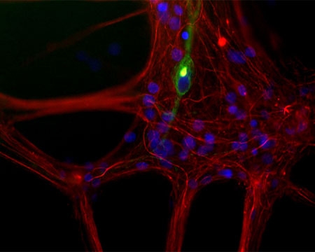
Mixed neuron/glia cultures from newborn rat brain stained with Mouse monoclonal antibody to Peripherin M-1400-500 (green) and Rabbit polyclonal antibody to Neurofilament Light R-1392-50 (red channel). A class of large neurons, like the one in the middle of this image, contain Peripherin, while the majority of neurons and their processes contain NF-L and not Peripherin. Interestingly, the Periperin positive cells often contain a cytoplasmic inclusion next to the nucleus which stains for both peripherin and NF-L, and so appears golden in this kind of image. The blue channel reveals the localization of DNA